Visual illusions are pretty cool! Whether it’s images of old ladies turning into young dames or colorful vortices seemingly revolving around themselves, people tend to be fascinated with the tricks that can be played on their brains with pretty simple 2D images.
One type of visual illusion are ‘bi-stable- images’, like the one below:
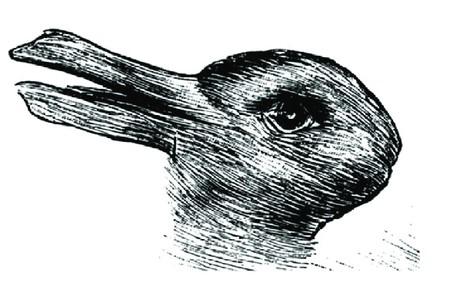
This is a relatively well-known bistable image called the ‘Jastow rabbit-duck’. As you may have noticed, it’s possible to see either a rabbit or a duck, but never both at the same time. Your perception switches between the two images, which is why it’s also called ‘bistable perception’.
Pretty awesome, right? Another kind of visual illusion out there is the ‘illusory contour’. Here, typically a geometric figure is seemingly observed even though no outline is given for such a figure. Let me demonstrate:
Most likely you see a second triangle on top of the outlined one, pointing downward, whose corners are defined by the three Pacman-like circles, like so:
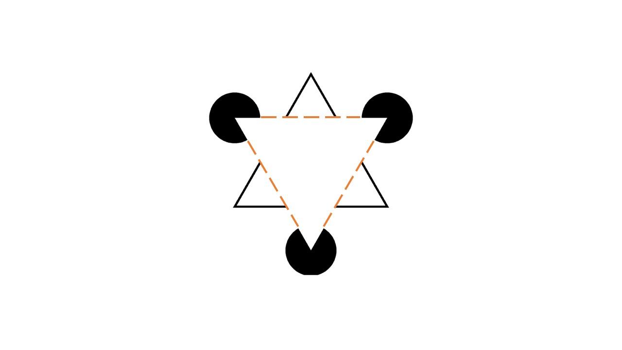
This triangle is our illusory contour, and this specific figure is called a ‘Kanizsa triangle’.
But how does that work in the brain exactly? Well, a paper by von der Heydt and colleagues investigated this in the visual cortex of macaque monkeys. Using microelectrodes that directly entered the animals’ brains, the researchers were able to record the electrical activity of individual neurons in the primary (V1) and secondary (V2) visual cortex*. They then showed an illusory contour to the monkeys and tried to record some neurons whose receptive field included the real, visible lines and some whose receptive fields included the illusory lines (like the ones outlined in orange above).
Never heard of ‘receptive fields’? Here’s a quick explanation: The receptive field of a neuron is basically the place in your field of view to which that particular neuron responds. Individual neurons in V1 only cover a tiny bit of your visual field, but together they cover all of it. That’s the same in V2 and other higher visual areas, but the receptive fields of the neurons actually get bigger, the higher up the visual area. So receptive fields in V2 for example are already a bit bigger than in V1, but not as big as in V4.
Then, when appropriate neurons were found in V1 and V2, the researchers moved the stimulus back and forth across the cells’ receptive fields. When doing this with a visible, non-illusory line, neurons in both V1 and V2 reacted with bursts of firing since the light in their receptive fields was changing during this movement. But what do you think happened for the neurons whose receptive fields only had moving illusory lines in them? Well, this is where it gets interesting: Neurons in V1 did not change their firing in response to the ‘lines’ moving, but many V2 neurons did. So, the neurons recorded from V2 responded as if the illusory line had actually been a real one! This may be the case since V1 seems to be responsive mostly to the orientation of seen stimuli and acts as a sort of ‘edge detector’ that tends to detect the outlines of things seen, but not much more**. V2 in contrast can already detect more complex visual features such as whether a figure is part of the foreground or background of an image. In general, the higher the visual area, the more complex the features it is there to detect. Thus, V2 neurons react to illusory contours which V1 neurons do not seem to ‘see’.
Alright, this concludes our foray into duck-rabbits, invisible triangles and our quirky visual system. Stay amazed, and ‘see’ you soon!
*If you’d like to learn more about the visual system of the brain, check out this article in our database: https://www.brainmatters.nl/en/database/visual-system/
**At least this is true early in the response to a stimulus (first 100ms). V1 also receives slightly delayed feedback from higher visual areas that broadens its functions + higher visual areas also receive (feedforward) information from lower ones such as V1, which are incorporated into their own responses!
Author: Melanie Smekal
References: