In addition to measuring brain activity, brain researchers can nowadays influence brain activity. We call this 'brain stimulation'. There are different forms of brain stimulation. The simplest form is simply to offer stimuli. After all, your brain is activated by all kinds of stimuli and even by your own thoughts. But here, of course, we are talking about a more direct form of brain stimulation.
Brain stimulation can be invasive or non-invasive. Invasive brain stimulation is, for instance, 'deep-brain stimulation', where electrodes are inserted directly into deep brain structures in patients with Parkinson's disease, among others.
Non-invasive brain stimulation takes place without opening the skull. It can be done in two ways: with magnetic pulses or with electric current. With magnetic pulses the brain is stimulated by TMS. With electricity stimulation can be done with a strong current as with ECT, or with low intensity as with TCS.
Author: Tom de Graaf (translated by Thomas von Rein)
Broca’s Area
Summary
Broca's area is important for spoken language. The famous patient of the discoverer of this area could only say "tan" because this area was damaged in him.
Function
Broca's area is used for speaking language and using grammar. This brain area is divided into two parts, the pars triangularis and the pars opercularis.
The pars triangularis is the anterior part of Broca's area, and important for various aspects of language, such as grammar and sentence structure. For example, this area helps with the sentence "he is punished by his mother. The structure of this sentence determines that "he" is being punished, and not the mother. When the pars triangularis is damaged, a person will understand this sentence as 'he punishes mother', which of course has a different meaning.
The pars opercularis is the posterior part of Broca's area, and especially important for speech. From this area, organs and muscles are controlled that make it possible to speak. The pars opercularis therefore has many connections to motor areas of the brain.
Location
In most individuals, Broca's area is primarily seen in the left hemisphere of the brain, on the surface of the frontal lobe. When Broca's area becomes damaged, however, it is possible that the same area in the right hemisphere may take over some functions.
Fact
Broca's area is named after the French physician Paul Pierre Broca. To this doctor came a patient who could only say "tantan. When the patient died, Broca examined his brain and found a lesion. The place where this damage was located was then named Broca's area, after its discoverer.
Patients
When Broca's area is damaged, it will be difficult to talk. These people prefer to use nouns, cannot apply grammar rules, or in the worst cases, cannot use meaningful words at all. This disorder is also known as Broca's aphasia.
Author: Myrthe Princen (translated by Thomas von Rein) Image: Marcel Loeffen
Basal Ganglia
The basal ganglia consist of a number of subcortical structures, the most important of which are the caudate nucleus, the putamen, and the globus pallidus. This group of structures has connections to very many parts of the cortex, but especially to the frontal lobe. The basal ganglia are therefore associated with movement planning, memory, emotion, reasoning and attention. Failure of the basal ganglia to function results in disorders such as Parkinson's and Huntington's disease, two diseases primarily related to the dysfunction of movements.
The caudate nucleus and putamen are known as the striatum, and this is the input nucleus of the basal ganglia. This means that they receive information from other parts of the brain, especially the cortex. The globus pallidus is the output nucleus of the basal ganglia, and so it receives information from the caudate nucleus and the putamen. The information is then transmitted to the thalamus. The thalamus then transmits this information to the motor and frontal areas in the cortex.
We have now explained the structures and connections of the basal ganglia. However, how these different structures work together is a whole different story, and not as easy as you might initially think. Logically, activity of the cortex would cause the basal ganglia to become active. This would then cause the thalamus to become active, and this would drive a movement. But this is not the case.
The part of the thalamus where the globus pallidus projects to is always active. This means that this area sends non-stop signals to the motor cortex. In daily life, of course, this cannot work because there are times when we have to sit still, or we only want to move a small part of our body. To counteract the fact that we are always moving, the globus pallidus has connections to the thalamus. The globus pallidus works with the neurotransmitter GABA, which has mainly inhibitory receptors. This means that the globus pallidus stops all activity of the thalamus (this is called inhibition).
When making a voluntary movement, signals from the cortex come to the caudate nucleus and the putamen. These two structures can then tell the globus pallidus to stop inhibiting some signals. So the globus pallidus is then "turned off" for a while, allowing these movements (the inhibition is thus inhibited, this is called disinhibition).
Author: Myrthe Princen (translated by Thomas von Rein)
Blood-Brain Barrier
The blood-brain barrier is the boundary between blood and fluid in the brain formed by endothelial cells and astrocytes. Endothelial cells are found in blood vessels, and throughout the body, they keep blood from flowing out of the blood vessels. Astrocytes are specialized glial cells, which help maintain the chemical environment of a neuron. The blood-brain barrier keeps bacteria and other water-soluble substances from entering the brain. Hormones can pass through the barrier, however, along with oxygen, carbon dioxide and glucose.
So, the blood-brain barrier is useful when it comes to protecting the brain. However, in some cases, it is also an additional hurdle when it comes to curing a disease in the brain; medicines cannot cross this barrier either. Therefore, in very severe cases, drugs are injected directly into the brain (kind of like with an epidural). The problem of the blood-brain barrier becomes especially apparent in Parkinson's disease, where there is too little dopamine in the brain. At first, it may seem simple to treat this: a dose of dopamine in a pill and any problem should be solved. However, dopamine is a substance that is not passed through the blood-brain barrier, and that’s why it’s so difficult to treat this disease.
Sometimes harmful substances also pass through the blood-brain barrier simply because they are too small to stop. Examples are hard drugs, such as cocaine and heroin, as well as caffeine and alcohol. This is also the reason why the cognitive effects of these substances become noticeable to the user pretty quickly.
Author: Myrthe Princen (translated by Melanie Smekal)
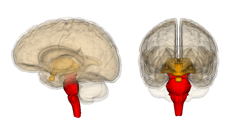
Brainstem
Summary The brainstem is important for many vital functions, such as breathing and heartbeat. The brainstem consists of the medulla, pons and midbrain.
Function The brain stem is important for vital functions, such as body temperature, heartbeat, breathing and blood pressure. It is also important for maintaining a sleep rhythm, crying, urinating, chewing and changing pupil size.
The brainstem consists of three parts; the medulla, the pons and the midbrain. The medulla is the part of the brainstem that is connected to the spinal cord. From here, signals are sent from the brain to the body, for example to the muscles to make a certain movement. In this way, the medulla is also involved in heartbeat, blood pressure, digestion and body temperature. The pons is the connection between the cerebellum and the cerebrum. The front part of the medulla is important for sending sensory information to the cerebellum. This mainly involves information about movement. The rear part of the pons helps with breathing, taste and sleep. The midbrain is involved in sensory and motor functions, visual and auditory reflexes, the pupillary reflex and hearing. The midbrain consists of the tectum and tegmentum. The tectum contains the superior and inferior colliculi, which control eye movements.
Location The brainstem is the connection between the cerebrum, cerebellum and spinal cord. The brainstem enters the skull through the opening at the base of the skull.
Fact The brainstem is the part of the brain that was first fully developed. This means that the first vertebrates already had a developed brain stem. The neurons that are found here are very small and have few long branches.
Patients Damage to the medulla of the brain stem can lead to paralysis. Body parts can also become insensitive to touch.
Author: Myrthe Princen (translated by Pauline van Gils)
Image: Marcel Loeffen
Manage Cookie Consent
To provide the best experiences, we use technologies like cookies to store and/or access device information. Consenting to these technologies will allow us to process data such as browsing behavior or unique IDs on this site. Not consenting or withdrawing consent, may adversely affect certain features and functions.
Functional
Always active
The technical storage or access is strictly necessary for the legitimate purpose of enabling the use of a specific service explicitly requested by the subscriber or user, or for the sole purpose of carrying out the transmission of a communication over an electronic communications network.
Preferences
The technical storage or access is necessary for the legitimate purpose of storing preferences that are not requested by the subscriber or user.
Statistics
The technical storage or access that is used exclusively for statistical purposes.The technical storage or access that is used exclusively for anonymous statistical purposes. Without a subpoena, voluntary compliance on the part of your Internet Service Provider, or additional records from a third party, information stored or retrieved for this purpose alone cannot usually be used to identify you.
Marketing
The technical storage or access is required to create user profiles to send advertising, or to track the user on a website or across several websites for similar marketing purposes.