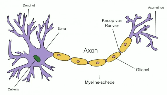
An axon is a thin spur of a neuron, and an essential component for the transmission of information. The beginning of the axon is found in the axon hillock, a bump on the cell body. The axon ends at the synapse, where communication with another cell is possible through the transfer of neurotransmitters. Sometimes many axons have to go to the same area in the brain, and they bundle together. These bundles of axons are known as nerves, except in the brain, where we call a bundle of axons a tract.
The main function of the axon is to conduct an action potential. This is an electrical pulse that is moved by charged ions entering and leaving the axon. In this way, an axon can transport signals over a very long distance. Some axons are more than a metre long. To transmit an action potential quickly over such a long distance, neurons work together with glial cells. These cells coat the axon with a substance called myelin, which provides insulation. This prevents the electrical charge in the axon from leaking out. To make it possible for the ions to enter the axon anyway, the myelin sheath is sometimes interrupted. These interruptions are called Ranvier's nodes.
At the nodes of Ranvier, positive ions can flow into the cell and continue an action potential. The resulting peak voltage then spreads through the myelin-wrapped section of the axon until it reaches the next node. There the whole story begins again. An action potential thus jumps, as it were, from one opening in the myelin sheath to the next. Due to this system, an action potential can reach a speed of 150 metres per second.
Author: Myrthe Princen (translated by Thomas von Rein ) Image: Marcel Loeffen
Auditory Cortex
Summary
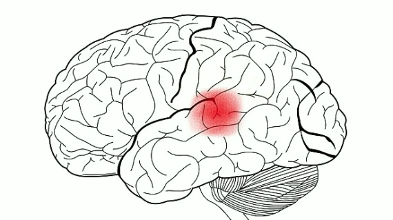
The auditory cortex is important for processing sounds. This involves identifying the pitch, volume and location of a sound. This involves using a topographical map.
Function
The auditory cortex is responsible for processing sounds. At the very beginning, processing of volume and pitch takes place. The auditory cortex is also responsible for localising a sound. The area is also important for processing speech.
Neurons in the auditory cortex respond best to a specific pitch (a specific frequency). Furthermore, neurons that respond to approximately the same frequencies are close together. This forms a topographical map of pitch, similar to the keys on a piano. This organisation is also called 'tonotopy'.
Location
The auditory cortex is located in the temporal lobe, just above the ears. Input to this area comes directly from the auditory portion of the thalamus.
Fact
Unlike the visual cortex, the auditory cortex develops very early. Small children are already very good at distinguishing different tones with low frequencies. However, the ability to localise tones is not fully developed until years later.
Patients
Damage to this area can cause people to be unaware of sounds. However, the sounds do enter the ear and there are reflex reactions to some sounds. Think of very loud sounds, which make you look up to see if there is any danger.
Author: Myrthe Princen (translated by Thomas von Rein) Image: Marcel Loeffen
Auditory System
The way in which the ear is shaped enables people to process sounds. Therefore, there are many different parts in the ear, each with its own function.
The structure of the ear is as follows:
Outer Ear
Pinna = auricle: collects sounds
The shape of the pinna helps us hear better when sounds are coming at us from the front. The shape of the pinna also enables us to localise sounds.
Auditory canal : reflects the sound waves
Middle Ear
Eardrum
Ossicles - Malleus = hammer - Incus = Anvil
Stapes = stirrup - The bottom of the stapes is called the footplate, and moves in and out of the oval window.
Inner Ear
Oval window
Cochlea = cochlea - Here, the vibrations of sound waves are converted into electrical signals that can be processed by neurons.
In order to transmit sound waves in the ear, the signals need to be amplified. This is because the cochlea is filled with fluid, which means that small vibrations are not passed on properly. The ossicles provide this amplification of the waves by acting as a lever. As a result, the signals entering the ear are amplified approximately 20 times between the eardrum and the oval window.
There are two muscles attached to the ossicles:
Tensor tympani muscle: attached to the hammer
Stapedius muscle: attached to the stirrup
When these muscles contract, the ossicles become much less movable, making it more difficult to amplify the signals. The contraction of these muscles is called the attenuation reflex, and aims to protect the auditory system.
The cochlea is a spiral-shaped structure that is filled with fluid inside. At the beginning of the cochlea we find two membranes, the round window and the oval window.
When a sound reaches the oval window, the basilar membrane starts to move. You can compare this to the swinging of a rope. When the frequency of a sound is high, it does not travel far across the basilar membrane. Low frequency sounds can travel to the end of the cochlea.
The basilar membrane contains auditory receptors. The place where these receptors are located is called the organ of Corti. On the organ of Corti there are hair cells. Each hair cell has about 100 protrusions, which are called stereocilia. When the basilar membrane moves, the stereocilia of the hair cells move too. The stereocilia of the different hair cells move as a unit, i.e. all at the same time and in the same direction. The hair cells are designed to depolarize when they move in one direction and hyperpolarize when they move in the other direction.
Hair cells have connections to spiral ganglion cells. These ganglion cells transmit their axons to the auditory nerve. The auditory nerve projects to the ventral and dorsal regions of the cochlear nucleus. From there, the ventral cochlear nucleus sends signals to the superior olive, and from the olive, the axons go to the inferior colliculi. From the inferior colliculi, the signals go to the medial geniculate nucleus (MGN) of the thalamus, and from there, of course, to the auditory cortex.
We can localise sound in different ways:
Time difference between ears (for sounds from 20 to 2000 Hz): it takes about 0.6 ms for a sound wave to travel from one side of the head to the other. If a sound comes directly from the right or directly from the left, it is thus received 0.6 ms earlier in one ear than in the other.
Intensity difference between ears (for sounds from 2000 to 20000 Hz): your head blocks part of the sound waves. When a sound comes directly from the right, there will be a difference in volume between the sound that enters the right ear and the sound that enters the left ear (in the right ear, the sound will be somewhat louder).
Comparing the input of the two ears (using the time difference or intensity difference) is useless to locate a sound vertically. However, it is possible for humans to localise sound in this way. This is possible because of the way our ear is shaped. The ear reflects sound waves in a specific way, depending on the angle at which the sound enters the ear.
Author: Myrthe Princen (translated by Thomas von Rein ) Image: Marcel Loeffen
Anterior Inferior Temporal Cortex
Summary
The anterior end of the temporal lobe is also called the anterior inferior temporal lobe, abbreviated as the aIT lobe. This area plays an important role in so-called semantic memories. It functions as a kind of collection center of different kinds of knowledge about objects and concepts.
Function
If you have a pork chop on your plate, you know it's called "pork chop”. Maybe you also know what animal it comes from, what it tastes like, what it feels like in your mouth. You know that you have to eat it, and also that that has to be done with a knife and fork. All these different kinds of information are stored in different places in the brain, and it is the aIT lobe that collects all this information, as it were, when it is needed.
Location
The temporal lobe is the piece of the brain that sits on either side of the brain, so to speak, behind the ears. At the very front extreme of this lobe is the anterior inferior temporal lobe, in line with the superior temporal gyrus and the medial temporal gyrus. It consists of about seven different sub-areas that are also found in monkey brains. However, there is one sub-area called "TG'' that exists only in humans.
Fact
The aIT lobe is usually one of the first areas to be affected during the development of Alzheimer's disease. This explains why people in the early stages of Alzheimer's sometimes have specific memory problems.
Patients
Patients with damage to this brain region often suffer from a condition called semantic amnesia. If they are asked to name a picture of a zebra, for example, they can often tell that it is a horse. Naming the picture requires little semantic knowledge about an object. But then they wonder why those horses have those crazy stripes and what they are for. Also, these patients have problems guessing what animal is meant by "an African animal with black and white stripes”. This is because it requires access to semantic memories.
Author: Myrthe Princen (translated by Thomas von Rein)
Anterior Cingulate Cortex
Summary
The anterior cingulate cortex (ACC) is involved in a variety of cognitive functions, but especially in tasks where cognitive conflicts must be overcome. This area is also involved in attention, and choice making.
Function
The ACC is associated with various cognitive functions, such as adapting to rewards, making decisions, empathy, and emotion. BA 32 is involved in error detection, among other things. Thus, the area becomes active the moment you notice that you have made an error in a task where you have to answer very quickly, for example.
A typical way to activate the cingulate cortex is by performing a task in which an error is elicited. You can think of the Stroop task, in which you have to name the color of the ink of words, instead of the words themselves. In this task, a conflict takes place between reading skills and the attempts to name the color of the ink.
Location
BA 32 is part of the dorsal anterior cingulate cortex. This is the anterior part of the cingulate cortex, an area folded around the corpus callosum.
Fact
Around the 2008 U.S. election, there was a lot of buzz around a study in London that reported a one-way relationship between the size of BA 32 and political orientation. The larger BA 32, the more left-wing a person's political affiliation would be. Without going into this particular study, it is advisable to be cautious with this type of result: one study does not make a truth.
Patients
Damage to this area is associated with a host of different symptoms, including not being able to detect errors, being inattentive, and difficulty with conflict tasks such as the Stroop task. People with attention disorder ADHD also show less activity in this brain region while performing cognitive tasks.
Author: Myrthe Princen (translated by Thomas von Rein)
Angular Gyrus
Summary
This area is involved in the functions of language, arithmetic and cognition. Some researchers think it is also involved in understanding and representing metaphors. Damage to the angular gyrus can cause Gerstmann syndrome.
Function
This area has several functions, all related to language, math and cognition. It has been found that people with a large BA 39 are good at math. In fact, this area is active in the exact calculation of certain sums. This area helps with this by retrieving mathematical facts from memory, and storing new knowledge again.
Some researchers think this area is important for understanding metaphors. Other researchers think there is no truth to this: these researchers refer to another area for this, namely the somatosensory cortex. Additionally, there are researchers who think that you can't attribute metaphors to one part of the brain, but that several areas are involved. Another example of how much there is still to discover!
Patients
Damage to this area can cause the development of Gerstmann syndrome. This disorder has four main symptoms, dysgraphia (difficulty writing), dyscalculia (difficulty doing math), finger agnosia (difficulty naming indicated fingers) and left-right disorientation.
Author: Myrthe Princen (translated by Thomas von Rein)
Adequate Stimuli
Adequate stimuli are the kinds of stimuli for which a sense's threshold is lowest. Each sense has a different adequate stimulus. For example, the photoreceptors in the retina of our eye are each most sensitive to a particular piece of the visual field. The hair cells in our ear respond to sound waves and each has a particular frequency of sound waves to which they respond best.
Author: Caroline Benjamins (translated by Anneke Terneusen)
Adaptation in the Retina
When you move from a brightly lit to an unlit/dark space, your eyes will need some time to get used to this new situation. At first, you will be able to perceive hardly any or no objects in the dark space. But after your eyes have had some time to get used to the new situation, you will be able to distinguish more objects. This adaptation process of your eyes is also called adaptation/habituation.
The process of adaptation in your retina is very different from neural adaptation. This is because the adaptation in your retina depends on the sensitivity of your photoreceptors. The adaptation process is entirely determined by the (maximum) sensitivity of the two photoreceptors located in the eye: the cones and rods. This process takes place in two stages: a so-called fast initial stage and a later, slower stage.
Both receptors begin to adjust their sensitivity as soon as the light goes out. However, the cones do this immediately with a much greater speed than the rods. Hence the so-called fast initial stage.
After a few minutes, the cones have reached their maximum sensitivity and they cannot adapt any further. Meanwhile, the rods have continued their adaptation process and reach the same level of sensitivity as the cones a few minutes after the cones have reached their maximum sensitivity. The rods, however, have not yet reached their maximum sensitivity and so the adaptation process continues for them. After another few minutes, the rods have also reached their maximum sensitivity. At this point, your eyes have adapted to the new situation.
Author: Caroline Benjamins (translated by Anneke Terneusen)
Anatomy
Many different terms are used to describe the brain. Some of these terms speak for themselves, and are not difficult to interpret. Other terms need a little more explanation, and so here is an overview of the most important terms, and what they mean.
To begin with, the brain is divided into several planes.
The horizontal plane divides the brain into a top and bottom half.
The sagittal plane divides the brain into a left and right side.
The coronal, or frontal, plane divides the brain into a front and back.
In addition, several terms are used to name the relative positions of different parts of the brain:
Anterior - in front of another part
Posterior - behind another part
Superior - above another part
Inferior - below another part
Lateral - further away from the center
Medial - more towards the center
Median - lying on the centre line
Proximal - near
Distal - far away from
Ipsilateral - on the same side of the brain
Contralateral - on the opposite side of the brain
There are still two important terms that remain when it comes to describing the brain. These concepts have ambiguous meanings, and therefore deserve some additional explanation. These are: ‘dorsal’ and ‘ventral’. These terms come from Latin, and mean "at the back" and "at the belly" respectively. In animals that walk on four legs, this is equivalent to 'the top of the body/head' and 'the bottom of the body/head', since the brain runs in a straight line looking from the spinal cord. In humans, however, the brain is tilted 90 degrees, and so here dorsal and ventral in the brain means something different than in the rest of the body.
Ventral
In the body: at the front
In the brain: at the bottom
Dorsal
In the body: at the back
In the brain: at the top
In addition to the general concepts mentioned above, anatomically the brain can also be divided into 52 different pieces. This division was made by a German neurologist named Korbinian Brodmann, and was based on differences in structure between the 52 areas. His division is still used to name the different parts of the brain. The different pieces are then designated by the characters BA (brodmann area) followed by a number.
Finally, the terms 'gyrus' (plural 'gyri') and 'sulcus' (plural 'sulci') are often used when naming anatomy. The 'coils' on the brain are the gyri, while the grooves between these coils are called sulci.
Author: Myrthe Princen (translated by Thomas von Rein)
Image: Marcel Loeffen
Do you want to know more content-wise about the anatomy of the brain? Check the Brain Basics articles here
Amygdala
The amygdala is a subcortical structure that is part of the limbic system. It is therefore involved in experiencing, processing and controlling various emotions. In addition, the amygdala is involved in many everyday functions of the brain.
The emotion most commonly associated with the amygdala is fear. Fear is an innate emotion, and already exists in babies when they unexpectedly hear a loud noise. The reaction that babies show is known as the startle reflex, and consists of blinking the eyes and contracting various muscles. During this reflex, activity is found in the amygdala. However, it would be too easy if the amygdala only became active during the emotion of fear. Activity is also seen in the amygdala when attention is focused on a particular emotion. There is a simple experiment to investigate this:
People are asked to evaluate well-known people as being 'good' or 'bad'. When asked to say how 'bad' a particular person is, the amygdala becomes active when seeing a person who is generally seen as bad (so, for example, when seeing a picture of Hitler). When asked to say how 'good' someone is, the amygdala does not become active when seeing a bad person, but becomes active when seeing a good person (e.g. when seeing a pop star).
In addition to the above reactions to emotions, the amygdala is also involved in certain expressions of emotion such as anger. When the amygdala is stimulated, it triggers a fight response. In this way, even the most peace-loving person can suddenly turn into a fighting creature.
The amygdala is also activated by the pons during dreaming. This is the reason why there are often many emotions in dreams. There is a theory that says that dreaming is nothing more than an attempt by the brain to process abnormal information. When you sleep you lie flat, in contrast to your posture during the rest of the day. This posture is interpreted as a position of flying or falling. According to this theory, this is the reason why so many people dream of free-falling or flying. This theory is called the activation-synthesis hypothesis.
Author: Myrthe Princen (translated by Pauline van Gils)
Attention controls the selective processing of different stimuli that are perceived at the same time. Thus, you need attention especially when there is a lot of input at once. By modulating attention toward a particular stimulus, we can process that stimulus better and faster. This is also the reason why people cannot read and listen well at the same time; attention only allows one of these functions to be performed properly.
The question is how the brain makes it possible for us to focus our attention in a way that is convenient for processing information. However, there is a problem that makes the study of attention a lot more difficult: the different types of attention.
To begin with, there is location-focused attention. This type of attention focuses on sounds or visual stimuli from a particular direction. For example, this occurs when you are at the bus stop waiting for the bus. Because you know from which direction the bus is coming, you will notice this bus faster than a bus in the opposite direction.
In addition to location-focused attention, there is attribute-dependent attention. Here you are specifically focused on properties that stimuli may have. When you go out on the town with a friend, you cannot process all the conversations that are within your hearing range. Instead, you mainly process the words and phrases that are characterized by the properties of your friend’s voice, so that you can respond to them appropriately.
Apparently, therefore, neurons in the sensory areas of the brain can adjust their response based on attentional processes. However, it is more difficult to find out exactly which brain region controls this attention. One area that is often associated with this is the pulvinar nucleus, an area in the posterior part of the thalamus. This association is made primarily on the basis of anatomical findings. Specifically, the pulvinar nucleus has many connections to the occipital, temporal, and parietal lobes. Problems with attention have also been found in several people with damage to the pulvinar nucleus. Yet there are also studies that conclude that this area is not the crucial area in directing attention. However, these studies do not offer an alternative as to how people focus their attention.
Author: Myrthe Princen (translated by Melanie Smekal)
Anatomy of the Eye
Our eye consists of several parts, as shown in this schematic image:
Below, we will give a brief description of the main parts of the eye that contribute to the visual process.
Pupil The pupil is the part of our eye that modulates the amount of light entering our eye. It is a hole in the middle of the iris. The light entering our eye does not reflect, which is why the pupil appears black.
Retina The retina is the part at the back of the eye that receives the entering rays of light. The retina is made up of several different cells, including photoreceptors.
Photoreceptors The retina contains about 126 million photoreceptors. These photoreceptors contain photopigments, which undergo decay when light falls on them. This creates an electrically charged current and allows signal transmission between neurons.
There are two types of photoreceptors:
Rods (+/-120 million) Rods are sensitive to little stimulation and thus become active when there is little light. These receptors are therefore mainly used at night.
Cones (+/- 6 million) The cones are sensitive to strong stimulation and are therefore mainly active during the day. This is because cones have photopigments that can be recharged very quickly. This allows them to recover quickly from light and to start decaying again. Cones are also essential for seeing colors.
Ganglion cells In addition to photoreceptors, the retina contains another type of cell called ganglion cells. These lie behind the photoreceptors and ensure that signals are projected to the brain. We distinguish two different types of ganglion cells:
Magno-ganglion cells: large cells, with large receptive fields
Parvo-Ganglion cells: small cells
All cells of the visual system have a receptive field. This is a concept that is difficult for some people to understand because it is not part of the cell. We describe the receptive field of a cell as the place in the visual space where light must be to make a particular cell active. You can also think of it in another way, as a place on the retina. In that case, the receptive field is the part of the retina where light has to fall to activate the cell further on in the brain.
Fovea/yellow spot In the center of the retina is the fovea, also called the yellow spot. This is where the light coming from the center of your vision falls. There are only cones in the fovea, which means that the center of the field of vision is also the place where you can see the most details.
Blind spot The location in the retina where the ganglion cells leave the eye and merge into the optic nerve has no photoreceptors. This means that light falling on this part of the retina cannot be processed. This area is also called the blind spot because you are "blind" to this area in your receptive field. This blind spot is "solved" in two ways. First of all, this spot is located in the peripheral part of your retina, the part where you cannot see very well in the first place. Secondly, the brain solves the phenomenon of the blind spot by copying the area in the receptive field that is just next to the blind spot. For example, when you look at a red sheet of paper, you do not suddenly see a black hole somewhere. The brain copies the image of the red area just next to your blind spot over the blind spot so that you will just see a whole red sheet of paper.
Author: Caroline Benjamins (adapted by Pauline van Gils) Illustration: Pauline van Gils
Manage Cookie Consent
To provide the best experiences, we use technologies like cookies to store and/or access device information. Consenting to these technologies will allow us to process data such as browsing behavior or unique IDs on this site. Not consenting or withdrawing consent, may adversely affect certain features and functions.
Functional
Always active
The technical storage or access is strictly necessary for the legitimate purpose of enabling the use of a specific service explicitly requested by the subscriber or user, or for the sole purpose of carrying out the transmission of a communication over an electronic communications network.
Preferences
The technical storage or access is necessary for the legitimate purpose of storing preferences that are not requested by the subscriber or user.
Statistics
The technical storage or access that is used exclusively for statistical purposes.The technical storage or access that is used exclusively for anonymous statistical purposes. Without a subpoena, voluntary compliance on the part of your Internet Service Provider, or additional records from a third party, information stored or retrieved for this purpose alone cannot usually be used to identify you.
Marketing
The technical storage or access is required to create user profiles to send advertising, or to track the user on a website or across several websites for similar marketing purposes.